Biotinylated Mouse TNFSF15 Protein
Catalog No: TNFSF15-MB001
- Species
- Mouse
- Expression System
- HEK293
- Tag
- His, Avi
- Activity
- Activity verified
Product overview
Recombinant Biotinylated Mouse TNFSF15 Protein is expressed in HEK293 cells with a His tag and Avi tag at the N-terminus. It contains amino acid residues Ala61-Leu252 (UniProt accession: Q5UBV8).
Product Details
- Molecular Aliases
- TL1A; VEGI-251; TNFSF15; TL1; VEGI; VEGI192A; VEGI251
- Protein Length
- Ala61-Leu252
- Expression System
- HEK293
- Theoretical Molecular Weight
- The protein has a predicted MW of 24.40 kDa. Due to glycosylation, the protein migrates to 28-38 kDa based on Bis-Tris PAGE result.
- Purity
- > 90% as determined by Bis-Tris PAGE
- Endotoxin
- Less than 1 EU per μg by the LAL method.
- Buffer / Formulation
- Lyophilized from 0.22μm filtered solution in PBS (pH 7.4). Normally 8% trehalose is added as protectant before lyophilization.
- State
- Lyophilized
- Storage Conditions
- -20 to -80°C for 12 months as supplied from date of receipt. -80°C for 3 months after reconstitution. Recommend to aliquot the protein into smaller quantities for optimal storage. Please minimize freeze-thaw cycles.
- Reconstitution Advice
- Dissolve the lyophilized protein in distilled water. Please refer to the Certificate of Analysis for detailed instructions.
Data Display
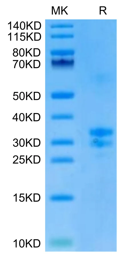
Biotinylated Mouse TNFSF15 on Bis-Tris PAGE under reduced condition. The purity is greater than 90%.
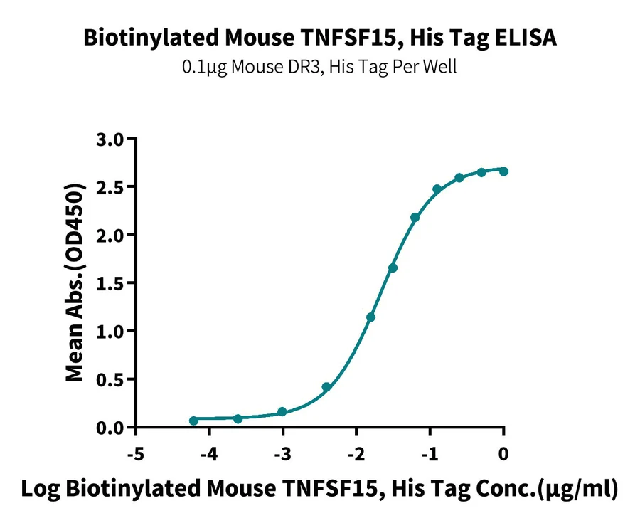
Immobilized Mouse DR3, His Tag at 1μg/ml (100μl/well) on the plate. Dose response curve for Biotinylated Mouse TNFSF15, His Tag with the EC50 of 21.4ng/ml determined by ELISA (QC Test).
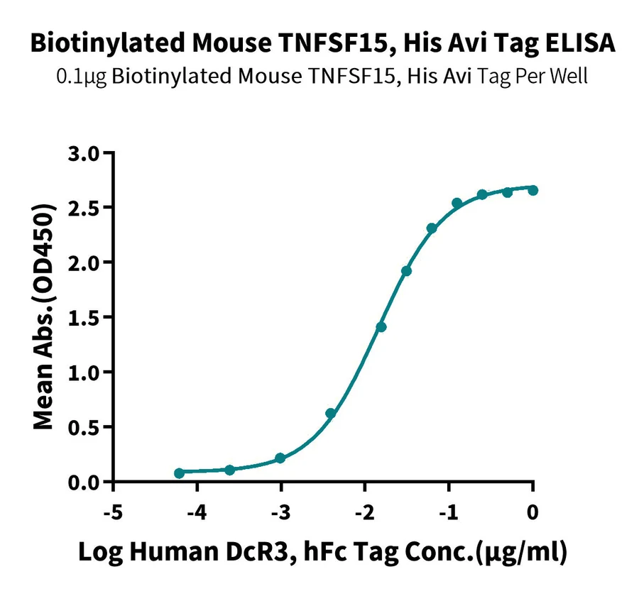
Immobilized Biotinylated Mouse TNFSF15, His Avi tag at 1μg/ml(100μl/well) on the streptavidin precoated plate(5μg/ml). Dose response curve for Human DcR3, hFc Tag with the EC50 of 14.4ng/ml determined by ELISA.
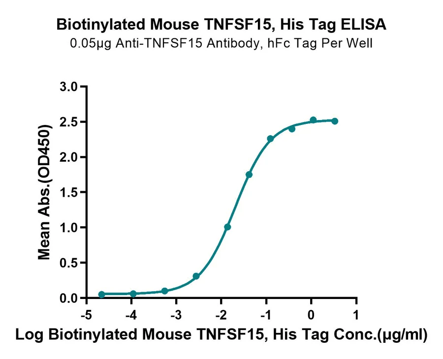
Immobilized Anti-TNFSF15 Antibody, hFc Tag at 0.5μg/ml (100μl/well) on the plate. Dose response curve for Biotinylated Mouse TNFSF15, His Tag with the EC50 of 20.5ng/ml determined by ELISA.
Background
TL1A and its functional receptor DR3 are members of the TNF/TNFR superfamilies of proteins. TL1A and DR3 are abundantly localized at inflamed intestinal areas of patients with IBD and mice with experimental ileitis or colitis and actively participate in the immunological pathways that underlie mucosal homeostasis and intestinal inflammation. Recently, an important role was demonstrated for TL1A/DR3 as potential mediators of intestinal fibrosis that is associated with the presence of gut inflammation.
References
- Valatas V, Kolios G, Bamias G. TL1A (TNFSF15) and DR3 (TNFRSF25): A Co-stimulatory System of Cytokines With Diverse Functions in Gut Mucosal Immunity. Front Immunol. 2019 Mar 27;10:583. doi: 10.3389/fimmu.2019.00583. PMID: 30972074; PMCID: PMC6445966.
Have questions about this product?
Our technical team can help with product selection, application questions and order support.